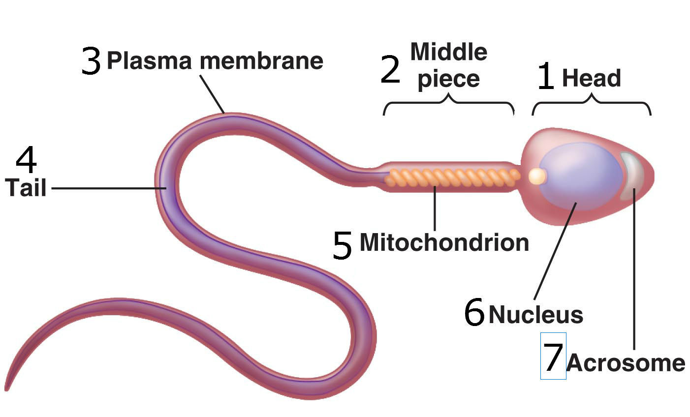
43 sperm cell diagram with labels
7th Grade Cells Search: Cells 7th Grade. A new study finds that 20 percent of third grade students have cell phones and 90 percent of them are online, while 83 percent of children in middle school have one share to facebook share to twitter Questions The _____ controls what enters and leaves the cell Victor, a seventh grader living in Fresno, CA, arrives for his first day of school Cell definition is - a ... DP Biology: Calculating Magnification and Size Activity 1 Calculating magnification of an image using it's scale bar. The three images below (click the eye to reveal) show a worked example of how to calculate sizes of cells organelles from electron micrographs step by step. Follow these steps carefully then complete the calculations on the worksheet.
A drug manufacturer has developed a time-release capsule...open 8 1Capacitation refers to the functional changes in sperm after they have been deposited in the female reproductive tract that enable them to fertilize a secondary oocyte. 2A morula is a solid ball of cells; a blastocyst consists of a rim of cells (trophoblast) surrounding a cavity (blastocyst cavity) and an inner cell mass. 3The blastocyst ...

Sperm cell diagram with labels
Structures of the ADGRG2-Gs complex in apo and ligand-bound forms ... the putative ligand-binding pocket in apo-adgrg2-fl was highlighted with red dashed square. d, vertical cross section of apo-adgrg2 (slate) disclosing a large cavity in the upper region of the 7tm... Labeled Diagram Of Developing Fetus In The Uterus Stock Vector Labeled Diagram Of Developing Fetus In The Uterus Stock Vector images that posted in this website was uploaded by Authtool2.britishcouncil.org. Labeled Diagram Of Developing Fetus In The Uterus Stock Vector equipped with a HD resolution 450 x 369.You can save Labeled Diagram Of Developing Fetus In The Uterus Stock Vector for free to your devices.. If you want to Save Labeled Diagram Of ... Flowering plant - Wikipedia Cross-section of a stem of the angiosperm flax: 1. pith, 2. protoxylem, 3. xylem, 4. phloem, 5. sclerenchyma ( bast fibre ), 6. cortex, 7. epidermis Angiosperm stems are made up of seven layers as shown on the right. The amount and complexity of tissue-formation in flowering plants exceeds that of gymnosperms.
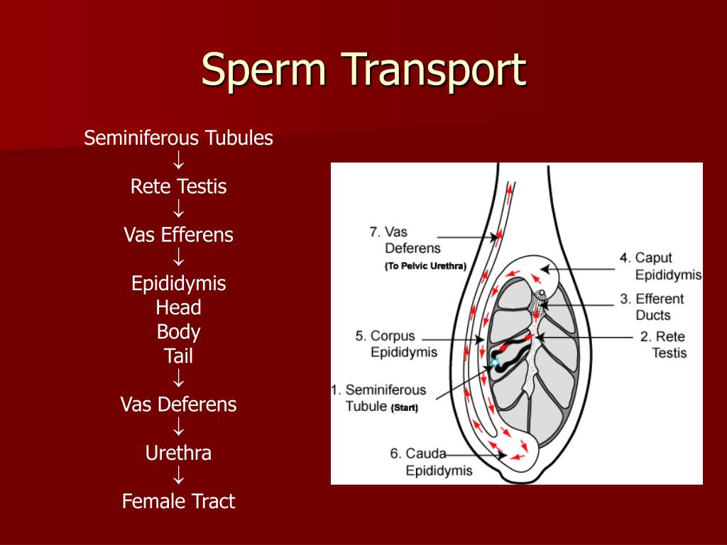
Sperm cell diagram with labels. 5 White Blood Cells Types and Their Functions - New Health Advisor 5. Basophils. Basophils are the least frequent type of white blood cell, with only 0-100 cells per mm 3 of blood. Basophils have large granules that perform functions that are not well known. They are very colorful when stained and looked at under the microscope, making them easy to identify. Frontiers | Identification of key genes and pathways in chronic ... Introduction. Chronic rhinosinusitis (CRS) is a disease characterized by chronic inflammation of the nasal cavity and sinus mucosa. The prevalence of CRS in the United States is 12.5%, whereas its prevalence in European countries and in China is 7% to 27% and 8%, respectively, thus resulting in high economic and social burdens (1-3).CRS mainly presents as phenotypes, namely, CRS without ... Urinogenital System (Theory) - Amrita Vishwa Vidyapeetham The process whereby primitive stem cell spermatogonia develop to form highly specialized spermatozoa is termed spermatogenesis. The testis is supplied by the internal spermatic artery, a highly coiled tortuous vessel. ... They are located along the route of the urethra as it relays sperm from the vas deferens out through the penis. The glands ... Biology Archive | August 16, 2022 | Chegg.com Biology archive containing a full list of biology questions and answers from August 16 2022.
centriolar satellite protein Cfap53 facilitates formation of the ... (A,B) Boxplots for sperm cell analysis showing the total number (horizontal lines) of sperm cells counted (A) and percentage of motile sperm cells (B) in spermatozoa derived from wild type ( n =7) and cfap53 −/− males ( n =7). Boxes indicate upper and lower quartiles and whiskers indicate upper and lower extremes. Brainstem: Definition, anatomy, parts, function | Kenhub Brainstem tectum, tegmentum and basal area (diagram) The tectum is the roof of the cavity while the tegmentum forms the ventral covering.The central cavity of the neural tube becomes the aqueduct of Sylvius, the fourth ventricle, and the central canal of the spinal cord.Therefore the tectum is the area dorsal to the aqueduct of Sylvius (in the midbrain) and fourth ventricle (at the pons ... Disposable Paper-Based Microfluidics for Fertility Testing Here, we review advancements in fabrication methods for paper-based microfluidic devices and their emerging fertility testing applications assessing sperm concentration, sperm motility, sperm DNA analysis, and other sperm functionalities, and provide a glimpse into future directions for paper-based fertility microfluidic systems. Graphical abstract Quiz Cell Labeling - pki.sicurezzalavoro.lombardia.it label the animal cell printout displaying top 8 worksheets found for - cell organelles labeling this is a basic illustration of a plant cell with major parts labeled note: a white cloudy solution should be seen mast cells can only be definitively recognized with special stains such as azure ii and toluoidine blue that identify the heparin storage …
Meiosis - Genome.gov In humans, body (or somatic) cells are diploid, containing two sets of chromosomes (one from each parent). To maintain this state, the egg and sperm that unite during fertilization must be haploid, with a single set of chromosomes. During meiosis, each diploid cell undergoes two rounds of division to yield four haploid daughter cells — the gametes. The Urinary System: Nephron & Urine Formation - Owlcation Main Structures of the Urinary System The main structures that comprise the urinary system are two kidneys (containing nephrons), two ureters, one bladder, one urethra, arteries and veins. The ureter connects the kidney to the bladder. The bladder is storage for urine. Urine is excreted to the outside of the body through the urethra. Kidney Basic Idea About IELTS examination Part 2; Listening You will need to complete labels on a plan (e.g. of a building), map (e.g. of part of a town) or diagram (e.g. of a piece of equipment). You can usually select your answers from a list on the question paper. This matching task assesses your ability to understand, for example, a description of a place, and to relate this to a visual representation. Antenatal Care Module: 3. Anatomy and Physiology of the Female ... Figure 3.1 Half section of the pelvic cavity showing the female reproductive organs, with the body facing to the left. You will learn more about the anatomical terms labelled in this diagram as this study session proceeds. Look carefully at Figure 3.1 for about two minutes, taking note of the position of the labelled structures.
draw and labeled diagram of mitochondria - Brainly.in
Sex Chromosome - Genome.gov Definition. 00:00. 00:29. A sex chromosome is a type of chromosome involved in sex determination. Humans and most other mammals have two sex chromosomes, X and Y, that in combination determine the sex of an individual. Females have two X chromosomes in their cells, while males have one X and one Y.
BTEC Applied Science Unit 1- Biology | Flashcards
Microscope Of Function And Quiz Parts Memorize information in a fun and engaging way Identify the parts that match with the description in the question microscope quiz These cells also accumulate at sites of infection, and the release of prostaglandins, serotonin and histamine help to increase blood flow to the area of damage, as part of the inflammatory response Parts of a ...
PPT - Male Anatomy PowerPoint Presentation, free download - ID:5970012
(PDF) Morpho-histology, endogenous hormone dynamics, and transcriptome ... by botanists and labels were present on the trees. ... elongate, reaches near the egg cell, releases sperm, and the sperm. ... structures, and cell mobility had the lowest unigenes, 186, 70,

NCERT Solutions for CBSE 12 Biology, Chapter 3 in PDF format
Female reproductive organs: Anatomy and functions | Kenhub Our labeled diagrams and quizzes on the female reproductive system are the best place to start. The uterus is supplied mainly by the uterine artery which arises from the internal iliac artery. The superior branch of the uterine artery supplies the body and fundus, while the inferior branch supplies the cervix.

The human egg cell explained for egg donors | Altrui
IJMS | Free Full-Text | Age-Dependent Variations in Functional Quality ... Increased male age is associated with a significant reduction in semen quality. Little is known about the sperm proteome changes resulting from the aging process. This study aimed to investigate the relationship between the functional quality and proteome of epididymal spermatozoa of dogs that were differing in age. The study was conducted on 30 male dogs that were divided into three age ...
Illustartion Showing Structure Sperm Cell Stock Vector 131979917 - Shutterstock
Proteomic Analysis of Murine Bone Marrow Very Small Embryonic-like Stem ... Supplementary Fig. 1 shows Venn diagrams of proteomes from BM-derived SSC-VSELs, HSCs, and MNCs at the steady-state conditions (left panel) and FSH + NAM VSELs (right panel). As shown, VSELs present a unique panel of protein expression that only partially overlapped with the proteome of HSCs and MNCs.

Free Ncert Solutions for 9th Class Science Tissues - Studyadda.com
Drosophila melanogaster - Wikipedia Drosophila melanogaster is a species of fly (the taxonomic order Diptera) in the family Drosophilidae.The species is often referred to as the fruit fly or lesser fruit fly, or less commonly the "vinegar fly" or "pomace fly". Starting with Charles W. Woodworth's 1901 proposal of the use of this species as a model organism, D. melanogaster continues to be widely used for biological research in ...
Post a Comment for "43 sperm cell diagram with labels"